Understanding How the Body Repairs Musculoskeletal Tissues
Every day, your body repairs countless microscopic injuries that occur through normal movement, exercise and the ageing process. Muscles, tendons, ligaments, cartilage, bone and joints all possess the capacity to heal, although each tissue repairs at a different rate and to a different extent.
Following injury, healing is not a single event but a carefully coordinated biological process involving inflammatory cells, platelets, blood vessels, connective tissue cells and chemical signalling molecules. Together, these components work to restore tissue structure and function.
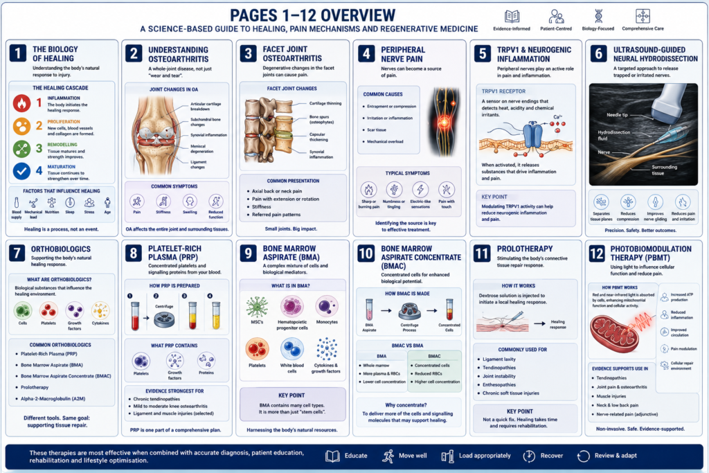
In many cases this process progresses successfully, allowing symptoms to resolve and normal activity to return. In some individuals,
however, healing becomes incomplete or disrupted, contributing to persistent pain, reduced function and chronic musculoskeletal disorders.
Understanding how tissues heal—and why healing sometimes fails—provides the foundation for modern musculoskeletal care.
The Normal Healing Response
Whether an injury occurs suddenly or develops gradually through repetitive loading, the body responds through a series of overlapping phases.
Phase 1 — Inflammation
Immediately after injury, the body initiates an inflammatory response. Although inflammation is often viewed negatively, it is an essential part of normal healing.
Specialised immune cells remove damaged tissue, control bleeding and release signalling molecules that coordinate the repair process. These signals attract additional cells required for healing and prepare the injured tissue for repair.
Phase 2 — Tissue Repair
As inflammation begins to settle, repair cells become active within the injured tissue.
Fibroblasts produce new collagen within tendons, ligaments and joint capsules, while other specialised cells participate in repairing muscle, bone and connective tissues. New blood vessels develop to improve nutrient delivery, and the extracellular matrix begins to reorganise.
During this phase the tissue gradually regains strength, although it remains vulnerable to excessive loading.
Phase 3 — Remodelling
Healing continues for many weeks and, in some tissues, many months.
New collagen fibres gradually reorganise along the direction of normal mechanical loading, becoming stronger and more functional. Progressive rehabilitation, appropriate exercise and gradual return to activity are essential during this stage to optimise recovery.
For many musculoskeletal injuries, the remodelling phase is where long-term improvements in strength, function and resilience occur.
When Healing Does Not Progress Normally
Not every injury follows an ideal healing pathway.
Repeated mechanical loading, persistent inflammation, ageing, metabolic factors and reduced tissue quality may interfere with the normal repair process.
Instead of progressing through healing, tissues may enter a cycle of ongoing inflammation, degeneration and incomplete repair. This may contribute to chronic tendon disorders, ligament laxity, osteoarthritis, persistent muscle injuries and some forms of chronic spinal pain.
Modern research increasingly recognises that many chronic musculoskeletal conditions involve changes affecting the entire biological environment of the tissue rather than damage to a single structure alone.
The Biology of Chronic Musculoskeletal Disorders
Persistent musculoskeletal pain is often more complex than a simple structural injury.
In conditions such as osteoarthritis and chronic tendinopathy, changes may occur within multiple tissues, including:
- Articular cartilage
- Synovial membrane
- Subchondral bone
- Tendons and ligaments
- Joint capsule
- Peripheral nerves
- Surrounding muscles and fascia
These tissues communicate continuously through cells, growth factors, inflammatory mediators and mechanical forces.
When this biological environment becomes disrupted, normal healing may become less efficient and symptoms may persist despite the original injury having occurred months or even years earlier.
The Importance of the Local Healing Environment
Successful tissue repair depends not only on the presence of healthy cells but also on the environment in which those cells function.
Factors that may influence tissue healing include:
- Persistent inflammation
- Mechanical overload
- Poor tissue quality
- Reduced blood supply
- Age-related biological changes
- Metabolic health
- Smoking
- Diabetes
- Nutrition
- Physical activity
Optimising these factors is often an important component of successful rehabilitation.
Supporting the Body’s Natural Healing Response
Modern musculoskeletal care increasingly focuses on creating the most favourable biological environment for tissue repair.
For many patients this begins with conservative treatment, including:
- Patient education
- Activity modification
- Progressive rehabilitation
- Exercise prescription
- Manual therapy where clinically appropriate
- Optimisation of general health
For carefully selected patients, additional procedures may also be considered as part of a comprehensive management plan.
These may include:
- Platelet-Rich Plasma (PRP)
- Bone Marrow Aspirate (BMA)
- Bone Marrow Aspirate Concentrate (BMAC)
- Prolotherapy
- Photobiomodulation Therapy (PBMT)
These approaches are commonly referred to as orthobiologic therapies. Rather than replacing damaged tissue directly, they aim to support or influence the local biological processes involved in tissue repair. Their role varies depending on the condition being treated, the quality of the available evidence and the individual patient’s circumstances.
Every Patient Heals Differently
No two injuries are identical, and no single treatment is appropriate for every patient.
The most suitable management plan depends on many factors, including:
- The underlying diagnosis
- The severity and duration of symptoms
- Tissue quality
- General health
- Functional goals
- Previous treatments
- Current scientific evidence
For this reason, treatment recommendations are always individualised following a comprehensive clinical assessment.
A Comprehensive Approach to Musculoskeletal Care
At Ashmore Osteopathic Group, treatment recommendations are based on understanding the underlying cause of your symptoms rather than focusing solely on pain.
Depending on your diagnosis, management may include education, rehabilitation, osteopathic care, diagnostic musculoskeletal ultrasound, image-guided procedures or orthobiologic therapies. These approaches are integrated into a personalised management plan designed to support recovery, improve function and help you return to your normal activities.
Continue Exploring
Understanding the biology of healing provides the foundation for many areas of modern musculoskeletal medicine.
You can learn more about:
- Understanding Osteoarthritis
- Facet Joint Osteoarthritis
- Peripheral Nerve Pain
- Neurogenic Inflammation and TRPV1
- Prolotherapy
- Platelet-Rich Plasma (PRP)
- Bone Marrow Aspirate (BMA)
- Bone Marrow Aspirate Concentrate (BMAC)
- Ultrasound-Guided Neural Hydrodissection
The information provided on this page is intended for general educational purposes only and should not be considered a substitute for an individual clinical assessment. Treatment recommendations are made following a comprehensive evaluation of your condition, current evidence and your individual healthcare needs.
page 2..
Understanding Osteoarthritis
Why Osteoarthritis Is More Than “Wear and Tear”
Osteoarthritis is the most common form of arthritis and one of the leading causes of musculoskeletal pain and disability worldwide. It can affect almost any synovial joint, including the knees, hips, shoulders, spine, hands and feet.
For many years, osteoarthritis was described simply as the gradual wearing away of joint cartilage. Modern research has shown that this explanation is incomplete. Osteoarthritis is now recognised as a condition affecting the entire joint, involving changes to cartilage, bone, the synovial lining, ligaments, joint capsule and surrounding muscles.
Although osteoarthritis becomes more common with age, it is not an inevitable consequence of ageing. Previous injury, repetitive mechanical loading, genetics, body weight, muscle strength, metabolic health and lifestyle all influence an individual’s risk of developing symptomatic osteoarthritis.
A Healthy Joint
Healthy joints are remarkable biological structures designed to provide smooth, pain-free movement while distributing mechanical loads during everyday activities.
A healthy synovial joint consists of:
- Articular cartilage, which provides a smooth, low-friction surface for movement.
- Subchondral bone, which supports the cartilage and absorbs mechanical forces.
- Synovium, a specialised lining that produces synovial fluid to lubricate and nourish the joint.
- Joint capsule and ligaments, which provide stability.
- Muscles and tendons, which control movement and reduce stress on the joint.
These structures function together as a single biological unit. When one component becomes damaged, the others often adapt, and over time these changes may contribute to osteoarthritis.
How Osteoarthritis Develops
Osteoarthritis usually develops gradually over many years.
Repetitive mechanical loading, previous injury or age-related biological changes may alter the balance between tissue repair and tissue breakdown within the joint.
Early changes often involve the cartilage, where small surface irregularities and loss of important structural molecules such as proteoglycans reduce the cartilage’s ability to resist compressive forces.
As these changes progress, the entire joint responds.
The synovial membrane may become inflamed (synovitis), the underlying subchondral bone remodels, bone marrow lesions may develop, and new bone (osteophytes) may form around the joint margins. Together, these biological and structural changes characterise osteoarthritis as a disease of the whole joint.
Why Osteoarthritis Can Become Painful
One of the most important discoveries in osteoarthritis research is that articular cartilage itself does not contain pain fibres.
Pain usually arises from other structures within and around the joint, including:
- The synovial membrane (synovitis)
- Subchondral bone
- Bone marrow lesions
- Joint capsule
- Ligaments
- Surrounding muscles and fascia
- Peripheral sensory nerves
This helps explain why some people with significant changes on X-rays experience little or no pain, while others with relatively mild imaging findings may have substantial symptoms.
Pain is influenced not only by structural changes but also by inflammation, joint mechanics, muscle function and the sensitivity of the nervous system.
Factors That Influence Osteoarthritis
Many factors contribute to the development and progression of osteoarthritis, including:
- Previous joint injury
- Repetitive occupational or sporting activities
- Age-related biological changes
- Family history and genetics
- Excess body weight
- Reduced muscle strength
- Joint instability
- Metabolic health
- Chronic low-grade inflammation
Not all of these factors can be modified, but many can be addressed through an individualised management plan.
Symptoms of Osteoarthritis
Symptoms vary between individuals and may include:
- Joint pain during or after activity
- Morning stiffness or stiffness after periods of rest
- Reduced joint movement
- Swelling or a feeling of joint fullness
- Clicking or grinding sensations (crepitus)
- Difficulty with everyday activities
- Reduced physical function
Symptoms often develop gradually and may fluctuate over time, with periods of improvement and exacerbation.
How Osteoarthritis Is Diagnosed
Diagnosis begins with a comprehensive clinical assessment.
This includes:
- A detailed medical history
- Assessment of symptoms and functional limitations
- Physical examination
- Evaluation of joint movement, strength and stability
Where clinically appropriate, imaging studies such as X-rays, MRI or diagnostic musculoskeletal ultrasound may provide additional information about joint structure and help exclude other conditions. Imaging findings are interpreted alongside the clinical assessment, as the severity of structural changes does not always reflect the severity of symptoms.
An Evidence-Informed Approach to Management
Management of osteoarthritis aims to reduce pain, improve function and support long-term joint health.
For most patients, treatment involves a combination of approaches rather than a single intervention.
Depending on the diagnosis and individual circumstances, management may include:
- Patient education
- Exercise therapy and progressive strengthening
- Weight management where appropriate
- Activity modification
- Osteopathic manual therapy where clinically indicated
- Diagnostic musculoskeletal ultrasound
- Image-guided musculoskeletal procedures
- Orthobiologic therapies, such as Platelet-Rich Plasma (PRP), when clinically appropriate and supported by current evidence
Treatment recommendations are individualised and developed through shared decision-making, taking into account your goals, overall health, current evidence and the potential benefits and limitations of each option.
Looking Beyond the Joint
Modern research continues to improve our understanding of osteoarthritis.
Rather than viewing it as simple cartilage wear, clinicians now recognise osteoarthritis as a condition involving complex interactions between mechanical loading, inflammation, tissue biology and the nervous system. This broader understanding has influenced the way musculoskeletal conditions are assessed and managed, with increasing emphasis on addressing the entire joint environment rather than focusing on cartilage alone.
Continue Exploring
Learn more about related topics:
- Facet Joint Osteoarthritis
- Knee Osteoarthritis
- Hip Osteoarthritis
- The Biology of Healing
- Platelet-Rich Plasma (PRP)
- Bone Marrow Aspirate (BMA)
- Bone Marrow Aspirate Concentrate (BMAC)
This information is provided for general educational purposes only and is not intended to replace an individual clinical assessment. Treatment recommendations are based on your diagnosis, clinical findings, current scientific evidence and your individual healthcare needs.
page 3…
Facet Joint Osteoarthritis (Facet Arthropathy)
Understanding Arthritis of the Spinal Facet Joints
The facet joints (also known as zygapophyseal joints) are small paired synovial joints located at the back of every spinal motion segment. Together with the intervertebral discs, they guide spinal movement, contribute to spinal stability and help distribute mechanical forces during everyday activities.
Like the knee, hip and shoulder, facet joints are true synovial joints. They contain articular cartilage, a synovial lining, a fibrous joint capsule and synovial fluid that allows smooth, low-friction movement.
Over time, injury, repetitive mechanical loading or age-related biological changes may lead to degeneration of these joints, resulting in facet joint osteoarthritis, also referred to as facet arthropathy or facet joint syndrome.
The Role of the Facet Joints
Every movement of the spine depends on the coordinated function of the intervertebral disc and the paired facet joints.
Together they:
- Guide normal spinal movement.
- Help maintain spinal stability.
- Distribute mechanical loads.
- Limit excessive movement that could damage the spine.
- Protect surrounding nerves and supporting structures.
The orientation of the facet joints differs throughout the spine, allowing each region to perform different movements.
Cervical Spine
The cervical facet joints are positioned in an oblique plane, allowing a wide range of movement including:
- Flexion
- Extension
- Rotation
- Side bending
This orientation makes the cervical spine the most mobile region of the vertebral column.
Lumbar Spine
The lumbar facet joints are orientated predominantly in the sagittal plane.
This arrangement allows:
- Flexion
- Extension
while limiting excessive rotation that could overload the lumbar intervertebral discs.
How Facet Joint Osteoarthritis Develops
Facet joint osteoarthritis usually develops gradually.
Repetitive mechanical loading, previous injury or degeneration of the intervertebral disc alters the normal mechanics of the spinal motion segment.
As disc height decreases, greater forces are transferred to the facet joints.
Over time this may result in:
- Cartilage degeneration.
- Synovitis (inflammation of the joint lining).
- Thickening of the joint capsule.
- Subchondral bone remodelling.
- Bone marrow lesions.
- Osteophyte formation.
- Enlargement of the facet joints (facet hypertrophy).
These changes occur progressively and may contribute to pain, stiffness and reduced spinal mobility.
Osteoarthritis Is a Disease of the Whole Joint
Current research recognises that facet joint osteoarthritis is not simply the result of cartilage wearing away.
Instead, it involves biological changes affecting the entire joint, including:
- Articular cartilage
- Synovial membrane
- Subchondral bone
- Joint capsule
- Ligaments
- Surrounding muscles
- Peripheral sensory nerves
These tissues communicate continuously through inflammatory mediators, growth factors and mechanical loading.
When this biological environment becomes disrupted, inflammation and tissue degeneration may become self-perpetuating, contributing to persistent spinal pain.
Why Facet Joints Become Painful
Healthy articular cartilage contains no pain fibres.
Pain associated with facet joint osteoarthritis usually arises from surrounding pain-sensitive structures, including:
- Inflamed synovium.
- Joint capsule.
- Subchondral bone.
- Periosteum.
- Supporting ligaments.
- Adjacent muscles and fascia.
- Peripheral sensory nerves.
This helps explain why the severity of imaging findings does not always correspond to the severity of symptoms.
Common Symptoms
Facet joint pain commonly presents as:
- Localised neck pain or lower back pain.
- Pain that increases with standing or walking.
- Pain aggravated by spinal extension (leaning backwards).
- Pain during spinal rotation.
- Morning stiffness.
- Stiffness after prolonged sitting.
- Reduced spinal mobility.
- Tenderness over the affected joints.
Pain may also be referred into the shoulder region, buttock or upper thigh without indicating nerve compression.
When Nerves Become Involved
Facet joint osteoarthritis does not usually cause numbness, tingling or muscle weakness by itself.
However, progressive degenerative changes may narrow the spaces through which spinal nerves travel.
These changes may include:
- Facet hypertrophy.
- Osteophyte formation.
- Thickening of supporting ligaments.
- Degenerative disc disease.
When spinal nerves become irritated or compressed, symptoms may include:
- Pain radiating into the arm or leg.
- Pins and needles.
- Numbness.
- Muscle weakness.
- Altered reflexes.
These neurological symptoms suggest nerve involvement and require further clinical assessment.
How Facet Joint Osteoarthritis Is Diagnosed
Diagnosis begins with a detailed clinical assessment.
This includes:
- Medical history.
- Physical examination.
- Assessment of spinal movement.
- Evaluation of joint loading patterns.
- Neurological examination where appropriate.
Imaging may also assist diagnosis.
Depending on the clinical findings, this may include:
- X-rays.
- CT scans.
- MRI.
- Diagnostic musculoskeletal ultrasound for selected surrounding soft tissues.
In selected patients, image-guided diagnostic injections may also help determine whether the facet joint is contributing significantly to symptoms.
An Evidence-Informed Approach to Management
Management aims to improve function, reduce pain and optimise spinal health.
Most patients benefit from a combination of conservative approaches, including:
- Patient education.
- Activity modification.
- Progressive rehabilitation.
- Exercise therapy.
- Osteopathic manual therapy where clinically appropriate.
Where symptoms persist despite appropriate conservative care, additional interventions may be considered for carefully selected patients.
Depending on the diagnosis, these may include:
- Ultrasound-guided musculoskeletal procedures.
- Prolotherapy.
- Platelet-Rich Plasma (PRP).
- Bone Marrow Aspirate (BMA) in selected circumstances.
Treatment recommendations are individualised and based on clinical assessment, current scientific evidence and shared decision-making.
Looking Beyond the Joint
Facet joint pain rarely occurs in isolation.
The surrounding muscles, ligaments, intervertebral discs and peripheral nerves all contribute to spinal function and may influence the development or persistence of symptoms.
Understanding these relationships allows treatment to focus not simply on the painful joint but on restoring the function of the entire spinal motion segment.
Continue Exploring
You may also be interested in:
- Understanding Osteoarthritis
- The Biology of Healing
- Cervicogenic Headache
- Peripheral Nerve Pain
- Platelet-Rich Plasma (PRP)
- Bone Marrow Aspirate (BMA)
- Prolotherapy
- Ultrasound-Guided Neural Hydrodissection
This information is intended for general educational purposes only and should not replace an individual clinical assessment. Treatment recommendations are based on the underlying diagnosis, current evidence and each patient’s individual circumstances.
page 4…
Understanding Peripheral Nerve Pain
The Role of Peripheral Nerves in Musculoskeletal Pain
When people think about musculoskeletal pain, they often focus on joints, muscles or tendons. While these structures commonly contribute to pain, the peripheral nervous system also plays an essential role in how pain develops, persists and resolves.
Peripheral nerves do much more than carry pain signals to the brain. They continually communicate with surrounding tissues, helping regulate sensation, movement, blood flow, inflammation and tissue repair. When a peripheral nerve becomes irritated or sensitised, it may contribute to persistent pain even when the original injury has largely healed.
Understanding the role of peripheral nerves has become an increasingly important part of modern musculoskeletal medicine and pain science.
What Are Peripheral Nerves?
Peripheral nerves connect the brain and spinal cord to the rest of the body.
They provide:
- Sensation from the skin, joints and muscles.
- Motor control to muscles.
- Communication between tissues and the nervous system.
- Regulation of local blood flow and tissue health.
Healthy peripheral nerves are designed to glide smoothly through surrounding muscles, fascia and connective tissues during normal movement. This process, known as neural gliding or neural excursion, allows nerves to accommodate changes in body position without excessive tension or compression.
How Peripheral Nerves Become Painful
Peripheral nerves may become irritated for many reasons, including:
- Acute injury.
- Repetitive mechanical loading.
- Chronic inflammation.
- Surgery.
- Scar tissue formation.
- Reduced tissue mobility.
- Compression within anatomical tunnels.
- Degenerative musculoskeletal conditions.
When the tissues surrounding a nerve become thickened or inflamed, the nerve may lose its normal ability to glide during movement. Increased mechanical stress, together with changes in the local biological environment, may lead to peripheral nerve sensitisation.
Rather than functioning normally, the affected nerve becomes more responsive to mechanical loading and may generate pain in response to movements or pressures that would not normally be painful.
Neurogenic Inflammation
Peripheral nerves are not passive structures.
When sensory nerves become irritated or sensitised, they release naturally occurring signalling molecules known as neuropeptides, including:
- Substance P
- Calcitonin Gene-Related Peptide (CGRP)
These molecules contribute to a process known as neurogenic inflammation.
Unlike the inflammatory response that occurs after an acute injury, neurogenic inflammation is driven by the nervous system itself. It may increase blood flow, alter communication between nerves and immune cells, influence surrounding connective tissues and amplify pain sensitivity.
Current research suggests that neurogenic inflammation may contribute to a variety of persistent musculoskeletal pain conditions.
Pain Is More Than Tissue Damage
One of the most important advances in pain science is the recognition that persistent pain does not always reflect ongoing tissue injury.
Following an injury, tissues may heal while the nervous system remains sensitised. In these situations, pain can continue because peripheral nerves have become mechanically or chemically hypersensitive rather than because tissue damage is progressing.
This helps explain why some people continue to experience pain despite relatively minor imaging findings, while others with more significant structural changes may have few symptoms.
Pain is influenced by the interaction between tissue health, biomechanics, inflammation and the nervous system.
Peripheral Nerve Entrapment
Certain peripheral nerves travel through narrow anatomical spaces where they may become mechanically irritated.
Common examples include:
- Median nerve (carpal tunnel syndrome)
- Ulnar nerve (cubital tunnel syndrome)
- Posterior interosseous nerve (lateral elbow pain)
- Suprascapular nerve (selected shoulder conditions)
- Greater and lesser occipital nerves (selected headache disorders)
- Superior cluneal nerves (selected causes of lower back pain)
- Inferior calcaneal (Baxter’s) nerve (selected heel pain conditions)
Entrapment does not always involve severe compression. Even relatively small reductions in nerve mobility or chronic irritation of surrounding tissues may contribute to persistent symptoms.
Recognising Peripheral Nerve Pain
Symptoms vary depending on the nerve involved but may include:
- Localised burning pain.
- Sharp or shooting pain.
- Tingling or altered sensation.
- Pain radiating away from the original site.
- Increased sensitivity to pressure.
- Symptoms aggravated by particular movements or postures.
These symptoms often overlap with tendon, ligament or joint disorders, making careful clinical assessment essential.
Assessment
Diagnosis begins with a comprehensive clinical assessment.
This may include:
- Medical history.
- Physical examination.
- Assessment of movement patterns.
- Neurological testing.
- Functional assessment.
- Diagnostic musculoskeletal ultrasound where clinically appropriate.
Although MRI and X-rays are valuable for many conditions, they do not always identify peripheral nerve irritation or dynamic nerve disorders. Clinical findings therefore remain an important part of diagnosis.
An Evidence-Informed Approach to Management
Management depends on the underlying diagnosis and the factors contributing to nerve irritation.
Treatment may include:
- Patient education.
- Activity modification.
- Progressive rehabilitation.
- Improving flexibility and strength.
- Osteopathic manual therapy where clinically appropriate.
- Optimising movement patterns.
- Management of contributing musculoskeletal disorders.
For carefully selected patients, ultrasound-guided procedures such as neural hydrodissection or perineural injection therapy may also be considered as part of a comprehensive management plan. These procedures are discussed in more detail elsewhere within this educational series.
Looking Beyond Pain
Peripheral nerves form an essential part of the body’s communication network. They influence not only sensation but also movement, inflammation and tissue health.
Recognising when a peripheral nerve is contributing to persistent musculoskeletal pain allows management to address the underlying biological and mechanical factors rather than focusing solely on the site where pain is experienced.
As our understanding of pain science continues to evolve, appreciation of the role of peripheral nerves has become an increasingly important component of evidence-informed musculoskeletal care.
Continue Exploring
Learn more about related topics:
- TRPV1 and Neurogenic Inflammation
- Ultrasound-Guided Neural Hydrodissection
- Perineural Injection Therapy
- Superior Cluneal Nerve Entrapment
- Cervicogenic Headache
- The Biology of Healing
This information is provided for general educational purposes only and should not replace an in
dividual clinical assessment. Treatment recommendations are based on your diagnosis, clinical findings, current scientific evidence and your individual healthcare needs.
page 5…
TRPV1, Neurogenic Inflammation & Chronic Pain
Understanding Why Some Nerves Continue to Generate Pain
Pain is an essential protective mechanism. When tissues are injured, specialised sensory nerves alert the brain to potential harm, encouraging us to protect the affected area while healing occurs.
In most cases, this protective system settles as tissues recover. In some individuals, however, sensory nerves remain unusually sensitive long after the original injury has occurred. This persistent nerve sensitisation may contribute to ongoing musculoskeletal pain even when structural healing is largely complete.
Modern pain research has identified several biological mechanisms that help explain this process. One of the most extensively studied involves a receptor known as Transient Receptor Potential Vanilloid 1 (TRPV1).
What Is TRPV1?
TRPV1 is a specialised protein found on many small pain-sensing (nociceptive) nerve fibres.
It functions as a molecular sensor, helping the nervous system detect potentially harmful stimuli, including:
- Heat
- Mechanical stress
- Increased tissue acidity
- Inflammatory chemicals released after injury
Under normal circumstances, TRPV1 plays an important protective role by warning the body of tissue damage and contributing to the normal healing response.
When TRPV1 Becomes Sensitised
Following injury or persistent mechanical loading, TRPV1 receptors may become increasingly responsive.
Rather than activating only when significant tissue damage occurs, sensitised receptors may respond to relatively minor mechanical or chemical stimuli.
This process is known as peripheral sensitisation.
As a result, movements or pressures that would normally be painless may begin to produce discomfort, while previously painful movements may become increasingly painful.
Neurogenic Inflammation
Sensitised peripheral nerves do more than transmit pain signals.
When activated, they release naturally occurring signalling molecules called neuropeptides, including:
- Substance P
- Calcitonin Gene-Related Peptide (CGRP)
These molecules contribute to neurogenic inflammation.
Unlike the inflammation associated with acute tissue injury, neurogenic inflammation originates from the nervous system itself. It influences communication between nerves, immune cells and surrounding connective tissues, contributing to increased pain sensitivity and changes within the local tissue environment.
Current research suggests that neurogenic inflammation may play a role in a range of chronic musculoskeletal disorders.
Why Pain Can Persist
One of the most important concepts in contemporary pain science is that persistent pain does not always indicate ongoing tissue damage.
Following an injury, tissues may repair while peripheral nerves remain sensitised.
This helps explain why:
- Some individuals continue to experience pain despite relatively minor findings on imaging.
- Pain may fluctuate even when structural changes remain stable.
- Mechanical loading that was previously well tolerated may become painful.
- Symptoms sometimes persist after tissues have substantially healed.
Pain is influenced by many interacting biological, mechanical and neurological factors rather than by structural changes alone.
Neurogenic Inflammation and Musculoskeletal Disorders
Research continues to investigate the contribution of peripheral nerve sensitisation and neurogenic inflammation in a variety of musculoskeletal conditions, including:
- Tendinopathy
- Ligament injuries
- Plantar heel pain
- Lateral elbow pain
- Shoulder pain
- Chronic spinal pain
- Peripheral nerve entrapment syndromes
- Cervicogenic headache
The degree to which neurogenic inflammation contributes varies between individuals and should always be interpreted within the broader clinical picture.
Why Conventional Anti-Inflammatory Medicines May Not Fully Address Nerve Pain
Many anti-inflammatory medications, such as non-steroidal anti-inflammatory drugs (NSAIDs), work by reducing prostaglandin production through inhibition of cyclooxygenase (COX) enzymes.
These medicines are often effective for acute inflammatory conditions.
Peripheral nerve sensitisation, however, involves additional biological pathways, including TRPV1 activation and the release of neuropeptides such as Substance P and CGRP.
Because these mechanisms differ, conventional anti-inflammatory medicines may provide only partial symptom relief for some forms of neuropathic or neurogenic pain. Management therefore often requires a broader approach directed at the underlying cause of nerve irritation.
Understanding the Biological Environment
Current research increasingly recognises that persistent pain reflects changes occurring within the local biological environment rather than damage to a single structure alone.
Mechanical loading, inflammation, connective tissues, blood supply, immune cells and peripheral nerves all influence one another.
For this reason, effective management often involves more than treating the site where pain is experienced. Addressing contributing biomechanical factors, improving tissue function and reducing ongoing mechanical irritation may all form part of a comprehensive treatment plan.
Where Does This Knowledge Lead?
Understanding peripheral nerve sensitisation has influenced the development of several evidence-informed treatment approaches.
Depending on the underlying diagnosis and individual circumstances, management may include:
- Education
- Progressive rehabilitation
- Activity modification
- Optimisation of biomechanics
- Manual therapy where clinically appropriate
- Management of contributing musculoskeletal disorders
For carefully selected patients, ultrasound-guided procedures designed to address mechanical irritation of peripheral nerves may also be considered. These procedures are discussed in the following pages of this educational series.
Key Messages
- TRPV1 receptors help detect potentially harmful mechanical, thermal and chemical stimuli.
- Persistent activation of TRPV1 may contribute to peripheral nerve sensitisation.
- Sensitised nerves release neuropeptides such as Substance P and CGRP that contribute to neurogenic inflammation.
- Chronic musculoskeletal pain often reflects interactions between tissues, inflammation and the nervous system rather than structural damage alone.
- Management should address the underlying biological and mechanical factors contributing to symptoms.
Continue Exploring
You may also be interested in:
- Ultrasound-Guided Neural Hydrodissection
- Perineural Injection Therapy
- Superior Cluneal Nerve Entrapment
- Cervicogenic Headache
- Facet Joint Osteoarthritis
- The Biology of Healing
This information is provided for educational purposes only and should not be interpreted as medical advice. Every patient requires an individual clinical assessment, and treatment recommendations are based on the underlying diagnosis, current scientific evidence and the patient’s individual circumstances.
page 6…
Ultrasound-Guided Neural Hydrodissection & Perineural Injection Therapy
Restoring the Mechanical Environment Around Peripheral Nerves
Healthy peripheral nerves are designed to move freely through the surrounding muscles, fascia and connective tissues as the body moves. This normal movement, known as neural gliding, allows nerves to accommodate changes in limb position without excessive tension or compression.
Following injury, repetitive mechanical loading, surgery or chronic inflammation, the tissues surrounding a nerve may become thickened, scarred or less mobile. These changes can restrict normal nerve movement, increase mechanical sensitivity and contribute to persistent pain.
For carefully selected patients, ultrasound-guided neural hydrodissection and perineural injection therapy may be considered as part of a comprehensive management plan.
Understanding Neural Hydrodissection
Neural hydrodissection is a minimally invasive ultrasound-guided procedure designed to gently separate the tissue planes surrounding an irritated peripheral nerve.
Using high-resolution musculoskeletal ultrasound, the affected nerve is identified and visualised throughout the procedure. A fine needle is then guided adjacent to the nerve under continuous real-time imaging, allowing a solution to be placed around the nerve without entering the nerve itself.
The injected solution gently separates surrounding connective tissues, creating additional space around the nerve and helping to restore its normal ability to glide during movement.
The procedure is performed with the aim of reducing mechanical irritation while protecting nearby blood vessels, tendons and other important anatomical structures.
Why Ultrasound Guidance Is Important
Peripheral nerves are often only a few millimetres in diameter and may lie close to arteries, veins, muscles and tendons.
High-resolution ultrasound allows the practitioner to:
- Identify the affected nerve.
- Assess the surrounding soft tissues.
- Visualise scar tissue or altered tissue planes.
- Guide the needle in real time.
- Monitor the spread of the injected solution.
- Avoid nearby blood vessels and other sensitive structures.
Ultrasound guidance improves procedural accuracy and assists safe placement of the injection.
What Is Perineural Injection Therapy?
Perineural injection therapy involves placing a solution immediately around an irritated peripheral nerve rather than injecting into the nerve itself.
The most commonly used solution is 5% dextrose, although the choice of injectate may vary depending on the clinical situation and current evidence.
Unlike prolotherapy, which uses higher concentrations of dextrose to stimulate connective tissue repair, low-concentration dextrose appears to have different biological effects.
Current research suggests that 5% dextrose may help reduce peripheral nerve sensitisation and influence neurogenic inflammation. Although the precise biological mechanisms continue to be investigated, this neuromodulatory effect is considered one of the leading explanations for the improvements reported by some patients following treatment.
When Might These Procedures Be Considered?
Ultrasound-guided neural hydrodissection or perineural injection therapy may be considered when clinical assessment suggests that a peripheral nerve is contributing to a patient’s symptoms.
Examples may include selected cases of:
- Peripheral nerve entrapment.
- Chronic nerve irritation.
- Persistent musculoskeletal pain associated with peripheral nerve sensitisation.
- Occipital neuralgia and selected cervicogenic headaches.
- Superior cluneal nerve irritation.
- Certain upper and lower limb nerve entrapment syndromes.
Not every patient with nerve-related pain is a suitable candidate. Treatment recommendations are individualised following a comprehensive assessment and consideration of appropriate conservative management.
A Comprehensive Assessment Comes First
Symptoms arising from peripheral nerves often resemble disorders affecting muscles, tendons, ligaments or joints.
For this reason, diagnosis begins with a comprehensive clinical assessment that may include:
- Medical history.
- Physical examination.
- Neurological assessment.
- Functional movement assessment.
- Diagnostic musculoskeletal ultrasound where clinically appropriate.
- Review of relevant imaging.
The goal is to identify the underlying contributor to pain before considering whether any procedure is appropriate.
What Happens During the Procedure?
Although every procedure is tailored to the individual, treatment generally involves:
- Identification of the affected nerve using ultrasound.
- Skin preparation using sterile technique.
- Placement of a fine needle adjacent to the nerve under continuous ultrasound guidance.
- Slow injection of the selected solution to separate the surrounding tissue planes.
- Immediate reassessment of nerve mobility and symptoms where appropriate.
The procedure is performed without injecting directly into the nerve.
What Does Current Research Show?
Research into ultrasound-guided neural hydrodissection and perineural injection therapy continues to evolve.
Clinical studies suggest that these procedures may improve pain and function for selected peripheral nerve conditions. Proposed mechanisms include:
- Reduction in mechanical irritation.
- Restoration of normal nerve gliding.
- Improvement of the local tissue environment.
- Modulation of peripheral nerve sensitisation.
- Reduction of neurogenic inflammation.
Responses vary between individuals, and further high-quality research continues to define the role of these procedures within evidence-based musculoskeletal care.
Rehabilitation Remains Essential
The procedure itself is only one component of treatment.
Long-term outcomes are influenced by addressing the factors that contributed to nerve irritation in the first place.
Depending on the diagnosis, rehabilitation may include:
- Progressive strengthening.
- Mobility exercises.
- Neural gliding exercises.
- Activity modification.
- Biomechanical assessment.
- Manual therapy where clinically appropriate.
- Ongoing clinical review.
Integrating these approaches helps support recovery and reduce the likelihood of recurrent symptoms.
Is This Treatment Suitable for Everyone?
No.
Ultrasound-guided neural hydrodissection and perineural injection therapy are not appropriate for every patient or every pain condition.
Recommendations are based on:
- The underlying diagnosis.
- Clinical assessment.
- Severity and duration of symptoms.
- Response to previous treatment.
- Current scientific evidence.
- Individual patient goals and preferences.
Shared decision-making is an important part of determining whether these procedures form an appropriate component of a personalised management plan.
Continue Exploring
Learn more about:
- Peripheral Nerve Pain
- TRPV1 and Neurogenic Inflammation
- Superior Cluneal Nerve Entrapment
- Cervicogenic Headache
- Prolotherapy
- Platelet-Rich Plasma (PRP)
- The Biology of Healing
This information is provided for educational purposes only and should not replace an individual clinical assessment. All procedures involve potential benefits, limitations and risks. Treatment recommendations are individualised and based on the underlying diagnosis, current scientific evidence and each patient’s clinical circumstances.
page 7…
Orthobiologics
Supporting the Body’s Natural Healing Response
Orthobiologics are biological substances used in the management of musculoskeletal disorders with the aim of supporting the body’s normal tissue repair processes.
Unlike joint replacement surgery or synthetic implants, orthobiologic treatments do not replace damaged tissue. Instead, they utilise naturally occurring biological components that are thought to influence the local healing environment within injured or degenerative tissues.
Most orthobiologic procedures use the patient’s own (autologous) blood or bone marrow, reducing the risk of immune rejection while introducing biological components that normally participate in tissue repair.
As research in regenerative medicine continues to evolve, orthobiologics have become an area of growing interest for selected tendon, ligament, muscle, cartilage and joint disorders. Their role continues to be refined as new clinical evidence emerges.
Why Does the Local Healing Environment Matter?
The body has an extraordinary ability to repair injured tissues.
In many acute injuries, this healing response progresses naturally through inflammation, tissue repair and remodelling, allowing normal function to return.
Some musculoskeletal conditions, however, become chronic.
Persistent mechanical overload, repeated injury, ageing, metabolic factors and long-standing inflammation may alter the local biological environment, making tissue repair less efficient.
Rather than simply affecting one structure, chronic musculoskeletal disorders often involve changes within the surrounding cells, blood vessels, inflammatory mediators and connective tissues that normally coordinate healing.
Modern regenerative medicine aims to better understand this biological environment and, where appropriate, support the body’s own repair mechanisms.
What Are Orthobiologics?
Orthobiologics are naturally derived biological preparations that contain varying combinations of cells, platelets, proteins and signalling molecules involved in normal tissue repair.
Depending on the procedure, these biological components may include:
- Platelets
- Growth factors
- Cytokines
- White blood cells
- Monocytes and macrophage precursors
- Mesenchymal stromal cells (MSCs)
- Haematopoietic stem and progenitor cells
- Extracellular signalling proteins
The biological composition differs considerably between procedures, which is why different orthobiologic treatments may be considered for different clinical situations.
Common Orthobiologic Procedures
Several orthobiologic procedures are currently used within musculoskeletal medicine.
These include:
Platelet-Rich Plasma (PRP)
PRP is prepared from a sample of the patient’s own blood and contains a concentration of platelets suspended within plasma.
Platelets release numerous growth factors and signalling molecules involved in the normal healing response and have been investigated across a range of tendon, ligament and joint conditions.
Bone Marrow Aspirate (BMA)
Bone marrow aspirate contains a naturally occurring mixture of biological cells and signalling molecules, including platelets, immune cells, mesenchymal stromal cells and haematopoietic progenitor cells.
Rather than replacing damaged tissue, these biological components are thought to influence the local tissue environment involved in healing.
Bone Marrow Aspirate Concentrate (BMAC)
Bone marrow aspirate concentrate is produced by processing bone marrow aspirate to increase the concentration of selected cellular components while reducing unwanted elements such as excess red blood cells and plasma.
The exact cellular composition varies according to the processing system used.
Alpha-2-Macroglobulin (A2M)
Alpha-2-Macroglobulin is a naturally occurring plasma protein currently being investigated for its potential ability to bind certain enzymes involved in cartilage breakdown.
Research in this area remains ongoing.
How Do Orthobiologics Work?
Although each orthobiologic differs in composition, they share a common objective.
Rather than acting as replacement tissue, they are intended to influence the biological environment surrounding injured structures.
Current research suggests these biological preparations may contribute to tissue repair through mechanisms such as:
- Cellular signalling.
- Modulation of inflammatory processes.
- Recruitment of repair cells.
- Collagen remodelling.
- Support of normal healing pathways.
The precise biological mechanisms remain an active area of scientific investigation and are likely to differ between individual therapies.
What Does Current Evidence Show?
The scientific literature investigating orthobiologics has expanded rapidly over the past decade.
Evidence is strongest for some applications of Platelet-Rich Plasma, particularly selected chronic tendinopathies and mild to moderate knee osteoarthritis.
For other orthobiologic procedures, including bone marrow aspirate and bone marrow aspirate concentrate, research remains promising but continues to evolve.
Outcomes vary depending on:
- The condition being treated.
- Disease severity.
- Patient selection.
- Rehabilitation.
- Preparation methods.
- Treatment protocols.
For these reasons, orthobiologic procedures should be considered within the context of the available evidence rather than as universal treatments for musculoskeletal disease.
Orthobiologics Are One Part of Comprehensive Care
Successful management of musculoskeletal disorders rarely depends on a single treatment.
Most patients benefit from a comprehensive approach that may include:
- Patient education.
- Activity modification.
- Progressive rehabilitation.
- Exercise therapy.
- Manual therapy where clinically appropriate.
- Optimisation of biomechanics.
- Weight management where appropriate.
- Image-guided procedures for selected conditions.
Where clinically appropriate, orthobiologic procedures may form one component of this broader management strategy.
Is Everyone a Candidate?
No.
Orthobiologic procedures are not suitable for every musculoskeletal condition or every patient.
Recommendations depend on:
- An accurate diagnosis.
- The severity and duration of symptoms.
- Current scientific evidence.
- Overall health.
- Previous treatments.
- Individual goals and expectations.
Every recommendation follows a comprehensive clinical assessment and shared decision-making process.
Continue Exploring
Learn more about:
- Platelet-Rich Plasma (PRP)
- Bone Marrow Aspirate (BMA)
- Bone Marrow Aspirate Concentrate (BMAC)
- Prolotherapy
- The Biology of Healing
- Understanding Osteoarthritis
Orthobiologic procedures continue to be investigated across a range of musculoskeletal conditions. Treatment recommendations are individualised following clinical assessment and discussion of the current scientific evidence, potential benefits, limitations and alternative management options.
Section 2 page 8…
Platelet-Rich Plasma (PRP)
Using the Body’s Own Biology to Support Tissue Repair
Platelet-Rich Plasma (PRP) is an orthobiologic treatment prepared from a small sample of a patient’s own blood. By processing the blood, platelets become concentrated within a reduced volume of plasma, creating a preparation that contains a higher concentration of platelets than is typically found in circulating blood.
Platelets are best known for their role in blood clotting, but they also contain numerous naturally occurring growth factors and signalling proteins that participate in the body’s normal response to tissue injury.
PRP has become one of the most extensively researched orthobiologic treatments in musculoskeletal medicine and is being investigated across a range of tendon, ligament, muscle and joint conditions.
What Is Platelet-Rich Plasma?
Whole blood is made up of several components:
- Red blood cells
- White blood cells
- Platelets
- Plasma
PRP is created by collecting a small blood sample and processing it using a specialised centrifuge. This separates the blood into its individual components, allowing a platelet-rich layer to be collected.
The resulting preparation contains a greater concentration of platelets than whole blood while retaining plasma proteins that also contribute to normal tissue repair.
The exact composition of PRP varies depending on the preparation system and clinical objectives.
Why Are Platelets Important?
Platelets contain microscopic storage granules that release biologically active molecules when activated.
These include a range of growth factors and signalling proteins involved in the body’s normal healing response, including factors associated with:
- Cell signalling
- Blood vessel formation
- Collagen production
- Tissue remodelling
- Cellular communication
Rather than replacing damaged tissue, PRP is thought to influence the biological environment in which healing occurs.
Research continues to investigate the precise mechanisms through which PRP interacts with injured tissues.
Leukocyte-Rich and Leukocyte-Poor PRP
Not all PRP preparations are the same.
Some preparation systems produce leukocyte-rich PRP, which contains higher numbers of white blood cells, while others produce leukocyte-poor PRP, where most white blood cells have been removed.
The choice of preparation depends on several factors, including:
- The tissue being treated.
- The underlying diagnosis.
- Current scientific evidence.
- Clinical experience.
- Treatment goals.
Research continues to evaluate which preparation may be more appropriate for different musculoskeletal conditions.
How Is PRP Prepared?
The preparation process generally involves:
- Collecting a sample of the patient’s own blood.
- Processing the blood in a centrifuge to separate its components.
- Isolating the platelet-rich fraction.
- Preparing the PRP for ultrasound-guided injection where clinically appropriate.
Because PRP is produced from the patient’s own blood (autologous), the risk of allergic reaction or immune rejection is very low.
Strict sterile technique is used throughout the preparation and injection process.
What Conditions Has PRP Been Studied For?
PRP has been investigated across a wide range of musculoskeletal conditions.
Examples include:
- Chronic tendinopathies
- Tennis elbow
- Achilles tendinopathy
- Patellar tendinopathy
- Rotator cuff tendinopathy
- Plantar fasciopathy
- Mild to moderate knee osteoarthritis
- Selected ligament injuries
- Certain muscle injuries
The strength of evidence varies between conditions. While some applications are supported by a growing body of research, others remain under investigation.
Treatment recommendations are based on the available evidence together with individual patient assessment.
What Does the Research Show?
PRP is among the most extensively studied orthobiologic therapies.
Systematic reviews and clinical guidelines suggest that PRP may improve pain and function for selected chronic tendon disorders and some patients with mild to moderate knee osteoarthritis.
For other conditions, evidence remains variable, and further high-quality research is continuing to define its role.
Responses differ between individuals and depend on factors such as:
- The diagnosis.
- Duration of symptoms.
- Severity of tissue changes.
- Preparation method.
- Injection technique.
- Rehabilitation following treatment.
For this reason, PRP should not be viewed as a universal treatment for musculoskeletal pain but rather as one option that may be considered for carefully selected patients.
Why Is Ultrasound Guidance Used?
Many musculoskeletal structures are small and lie close to important nerves, blood vessels and adjacent tissues.
High-resolution musculoskeletal ultrasound allows the treating practitioner to:
- Confirm the target structure.
- Visualise tendons, ligaments, joints and surrounding soft tissues.
- Guide needle placement in real time.
- Monitor delivery of the PRP.
- Avoid nearby anatomical structures.
Ultrasound guidance helps improve procedural accuracy and ensures the biological preparation is delivered to the intended anatomical location.
PRP Is Only One Part of Treatment
The injection itself is only one component of care.
Successful outcomes depend on addressing the underlying mechanical and biological factors contributing to the condition.
Depending on the diagnosis, a comprehensive treatment plan may include:
- Patient education.
- Activity modification.
- Progressive rehabilitation.
- Strengthening exercises.
- Manual therapy where clinically appropriate.
- Biomechanical assessment.
- Ongoing clinical review.
Integrating rehabilitation with orthobiologic treatment is considered an important part of evidence-informed musculoskeletal care.
Is PRP Suitable for Everyone?
No.
PRP is not appropriate for every patient or every musculoskeletal condition.
Suitability depends on:
- The underlying diagnosis.
- Stage of the condition.
- General health.
- Previous treatments.
- Current medications.
- Patient goals.
- Current scientific evidence.
A comprehensive assessment helps determine whether PRP forms an appropriate part of an individualised management plan.
Frequently Asked Questions
Is PRP the same as stem cell therapy?
No.
PRP is prepared from blood and primarily contains concentrated platelets and plasma proteins. It does not contain the cellular composition found in bone marrow aspirate or bone marrow aspirate concentrate.
Is PRP surgery?
No.
PRP is a minimally invasive injection procedure performed without surgery.
Can PRP replace rehabilitation?
No.
Exercise therapy and rehabilitation remain fundamental components of managing most musculoskeletal conditions. PRP is considered an adjunct to, rather than a replacement for, a comprehensive rehabilitation program.
How many treatments are required?
The number of treatments varies according to the condition being managed, its severity and the individual’s response to treatment. Recommendations are made following clinical assessment and review.
Continue Exploring
You may also be interested in:
- Orthobiologics
- Bone Marrow Aspirate (BMA)
- Bone Marrow Aspirate Concentrate (BMAC)
- Prolotherapy
- The Biology of Healing
- Understanding Osteoarthritis
Platelet-Rich Plasma (PRP) is one of several orthobiologic procedures used in musculoskeletal medicine. Recommendations are individualised following clinical assessment and discussion of the available scientific evidence, potential benefits, limitations and alternative management options.
page 9…
Bone Marrow Aspirate (BMA)
Understanding the Biology of Bone Marrow
Bone Marrow Aspirate (BMA) is an orthobiologic procedure that uses a small sample of a patient’s own bone marrow. Bone marrow is a naturally occurring biological tissue that contains a diverse population of cells and proteins involved in blood formation, immune regulation and the body’s normal tissue repair processes.
Unlike Platelet-Rich Plasma (PRP), which is derived from blood, bone marrow contains a broader range of naturally occurring cellular components. For this reason, Bone Marrow Aspirate is being investigated as an orthobiologic option for selected musculoskeletal conditions.
Rather than replacing damaged tissue, BMA aims to support the biological environment involved in normal healing.
What Is Bone Marrow?
Bone marrow is the soft tissue found within many bones of the body.
Its primary role is the continuous production of blood cells throughout life, but it also contains a complex mixture of biological cells that participate in immune function, inflammation and tissue repair.
Bone marrow contains:
- Mesenchymal stromal cells (MSCs)
- Haematopoietic stem and progenitor cells
- Monocytes and macrophage precursors
- Platelets
- White blood cells
- Growth factors
- Cytokines
- Extracellular signalling proteins
These components work together within a highly regulated biological environment.
What Is Bone Marrow Aspirate?
Bone Marrow Aspirate is obtained by withdrawing a small volume of liquid bone marrow using a specialised aspiration needle under sterile conditions.
The procedure is most commonly performed from the posterior iliac crest (back of the pelvis) because this location provides safe access to bone marrow while minimising discomfort.
The aspirated bone marrow is then prepared for clinical use according to the intended procedure.
Depending on the treatment plan, the aspirate may be used directly or further processed to produce Bone Marrow Aspirate Concentrate (BMAC).
Bone Marrow Is More Than “Stem Cells”
The term “stem cell therapy” is frequently used in popular media but oversimplifies the biology of bone marrow.
Mesenchymal stromal cells represent only a very small proportion of the cells naturally present within bone marrow.
The majority of bone marrow consists of:
- Blood-forming cells.
- Immune cells.
- Platelets.
- Plasma proteins.
- Monocytes.
- Haematopoietic progenitor cells.
- Other supporting stromal cells.
Current research suggests that communication between these different cell populations may be just as important as the presence of any individual cell type.
For this reason, many researchers now describe bone marrow procedures as cell-based orthobiologic therapies rather than simply “stem cell treatments.”
Why Are Mesenchymal Stromal Cells Important?
Mesenchymal stromal cells (MSCs) have attracted considerable scientific interest because they are capable of producing a wide range of biologically active signalling molecules.
Rather than acting primarily by replacing damaged tissue, current evidence suggests that MSCs exert much of their influence through paracrine signalling.
This means they release proteins and extracellular vesicles that communicate with surrounding cells and may influence:
- Inflammatory responses.
- Immune regulation.
- Blood vessel formation.
- Collagen remodelling.
- Cellular communication.
- Tissue repair processes.
Research into these mechanisms continues to evolve.
The Importance of the Bone Marrow Microenvironment
Bone marrow functions as a biological ecosystem.
Its therapeutic potential is thought to arise from interactions between multiple cell populations rather than from any single cell type.
Scientists increasingly recognise that:
- Immune cells influence healing.
- Platelets provide signalling proteins.
- Monocytes contribute to tissue repair.
- Stromal cells coordinate cellular communication.
- Growth factors regulate tissue responses.
This complexity helps explain why bone marrow continues to be an active area of regenerative medicine research.
What Conditions Is BMA Being Investigated For?
Bone Marrow Aspirate has been investigated across a range of musculoskeletal conditions, including selected cases of:
- Osteoarthritis
- Tendon disorders
- Ligament injuries
- Cartilage lesions
- Bone marrow lesions
- Delayed tissue healing
The strength of evidence varies considerably between conditions, and recommendations should always be based on individual assessment and the current scientific literature.
What Does Current Evidence Show?
Research into Bone Marrow Aspirate is expanding rapidly.
Early clinical studies suggest that BMA may improve pain and function in selected musculoskeletal conditions. However, study methods, preparation techniques and treatment protocols vary considerably.
As a result, further high-quality clinical trials are required before the role of Bone Marrow Aspirate can be fully defined for many conditions.
Current evidence supports careful patient selection and integration with comprehensive rehabilitation rather than routine use for all musculoskeletal disorders.
Why Is Ultrasound Guidance Important?
Many bone marrow procedures are combined with ultrasound-guided musculoskeletal injections.
Ultrasound allows the treating practitioner to:
- Visualise the target tissue.
- Identify surrounding anatomical structures.
- Guide needle placement in real time.
- Deliver the biological preparation accurately.
- Minimise unnecessary tissue trauma.
Image guidance forms an important part of modern orthobiologic practice.
BMA Is Part of Comprehensive Care
Bone Marrow Aspirate is not intended to replace rehabilitation or other evidence-informed treatments.
Depending on the diagnosis, management may also include:
- Patient education.
- Progressive exercise therapy.
- Activity modification.
- Manual therapy where clinically appropriate.
- Biomechanical assessment.
- Weight management where appropriate.
- Ongoing clinical review.
Where appropriate, BMA may be considered as one component of an individualised management strategy.
Frequently Asked Questions
Is Bone Marrow Aspirate the same as stem cell therapy?
No.
Bone marrow contains a wide variety of naturally occurring cells. Mesenchymal stromal cells represent only one small component of this complex biological tissue.
Is BMA the same as BMAC?
No.
Bone Marrow Aspirate (BMA) is the freshly aspirated bone marrow. Bone Marrow Aspirate Concentrate (BMAC) is produced by processing BMA to increase the concentration of selected cellular components while reducing unwanted elements such as excess red blood cells and plasma.
Is bone marrow taken from the spine?
No.
Bone marrow is most commonly collected from the posterior iliac crest (the back of the pelvis), which provides safe access to the marrow cavity.
Does BMA replace surgery?
Not necessarily.
Management decisions depend on the diagnosis, severity of the condition, patient goals and current scientific evidence. For some conditions, surgery remains the most appropriate treatment, while for others, non-surgical management may be considered.
Continue Exploring
Learn more about:
- Bone Marrow Aspirate Concentrate (BMAC)
- Platelet-Rich Plasma (PRP)
- Orthobiologics
- Prolotherapy
- The Biology of Healing
- Understanding Osteoarthritis
Bone Marrow Aspirate (BMA) is an orthobiologic procedure that continues to be investigated across a range of musculoskeletal conditions. Treatment recommendations are individualised following comprehensive assessment, discussion of current scientific evidence, and consideration of potential benefits, limitations and alternative treatment options.
page 10…
Bone Marrow Aspirate Concentrate (BMAC)
Understanding Why Bone Marrow Is Sometimes Concentrated
Bone Marrow Aspirate Concentrate (BMAC) is an orthobiologic preparation produced by processing a patient’s own bone marrow aspirate using a specialised centrifugation system.
Rather than creating a different biological product, centrifugation changes the composition of the original bone marrow aspirate by increasing the concentration of selected cellular components while reducing others, such as excess red blood cells and plasma.
The aim is to produce a preparation enriched with naturally occurring cells and signalling molecules that participate in the body’s normal tissue repair processes.
BMAC represents one of several orthobiologic options currently being investigated for selected musculoskeletal conditions.
What Is the Difference Between BMA and BMAC?
Bone Marrow Aspirate (BMA) is the freshly collected liquid bone marrow obtained during aspiration.
Bone Marrow Aspirate Concentrate (BMAC) is created by processing that aspirate in a centrifuge.
This process does not create new cells or alter their biological function. Instead, it separates the aspirate into layers according to cell density, allowing selected components to be collected in a smaller volume.
In simple terms:
- BMA is the original biological sample.
- BMAC is a concentrated preparation of selected naturally occurring components from that sample.
Why Concentrate Bone Marrow?
Fresh bone marrow contains:
- Red blood cells
- Plasma
- Platelets
- White blood cells
- Monocytes
- Mesenchymal stromal cells (MSCs)
- Haematopoietic stem and progenitor cells
- Cytokines
- Growth factors
During centrifugation, the concentration of several of these biologically active components increases while unnecessary bulk, particularly excess red blood cells, is reduced.
The goal is to deliver a preparation with a greater concentration of cells and signalling molecules within a smaller injection volume.
What Does BMAC Contain?
Although the exact composition depends on the preparation system used, Bone Marrow Aspirate Concentrate typically contains:
- Platelets
- Monocytes
- White blood cells
- Mesenchymal stromal cells
- Haematopoietic stem and progenitor cells
- Endothelial progenitor cells
- Cytokines
- Growth factors
- Extracellular signalling proteins
These cells and proteins interact through complex biological signalling pathways that continue to be investigated in regenerative medicine research.
Why Reduce Red Blood Cells?
One purpose of concentration is the reduction of excess red blood cells.
Red blood cells are essential for oxygen transport within the circulation, but they are not thought to contribute significantly to the biological objectives of orthobiologic procedures.
Removing much of this volume allows a greater proportion of biologically active cellular components to be delivered within a smaller injection.
The degree of red blood cell reduction varies according to the processing system used.
Mesenchymal Stromal Cells Are Only One Part of BMAC
Much public discussion surrounding BMAC focuses on mesenchymal stromal cells (MSCs).
Although MSCs remain an important area of research, they represent only one component of a much more complex biological preparation.
Current evidence suggests that interactions between:
- Platelets
- Monocytes
- Immune cells
- Stromal cells
- Growth factors
- Cytokines
may collectively influence the biological environment involved in tissue repair.
Increasingly, regenerative medicine is viewed as a process of coordinated cellular communication rather than the action of a single cell type.
How Is BMAC Prepared?
The preparation process generally involves:
- Bone marrow aspiration under sterile conditions.
- Placement of the aspirate into a specialised centrifuge.
- Separation of the aspirate into its component layers.
- Collection of the concentrated cellular fraction.
- Ultrasound-guided delivery where clinically appropriate.
Preparation systems differ, meaning the final biological composition of BMAC may vary between devices and protocols.
What Conditions Is BMAC Being Investigated For?
Bone Marrow Aspirate Concentrate has been investigated for selected musculoskeletal conditions, including:
- Mild to moderate osteoarthritis
- Cartilage defects
- Tendon disorders
- Ligament injuries
- Bone marrow lesions
- Selected degenerative musculoskeletal conditions
The available evidence continues to evolve, and outcomes vary according to the condition being treated, patient selection, rehabilitation and preparation methods.
What Does Current Research Show?
Clinical research into BMAC has expanded considerably over recent years.
Early studies suggest that BMAC may improve pain and function in carefully selected patients with certain musculoskeletal disorders. However, study quality, treatment protocols and preparation techniques remain variable.
At present, further high-quality clinical trials are required to better define:
- Which conditions respond best.
- Optimal preparation methods.
- Appropriate patient selection.
- Long-term clinical outcomes.
For these reasons, BMAC should be considered within the context of evidence-informed musculoskeletal care rather than as a universal treatment.
Why Is Ultrasound Guidance Used?
Following preparation, BMAC is commonly delivered using ultrasound guidance where appropriate.
Real-time imaging allows the practitioner to:
- Identify the target tissue.
- Visualise surrounding anatomy.
- Guide needle placement accurately.
- Monitor delivery of the preparation.
- Avoid nearby nerves, blood vessels and tendons.
Ultrasound guidance supports accurate placement of the orthobiologic preparation while improving procedural precision.
BMAC Is One Part of a Comprehensive Treatment Plan
Like all orthobiologic procedures, BMAC is not a stand-alone treatment.
Successful management depends on addressing the underlying causes of symptoms through a comprehensive approach that may include:
- Patient education
- Progressive rehabilitation
- Activity modification
- Strength and conditioning
- Manual therapy where clinically appropriate
- Biomechanical assessment
- Ongoing clinical review
Where clinically appropriate, BMAC may be integrated into this broader management strategy.
Frequently Asked Questions
Is BMAC better than PRP?
Not necessarily.
PRP and BMAC are different orthobiologic preparations with different biological compositions. The choice depends on the condition being treated, current scientific evidence and individual patient assessment.
Is BMAC the same as stem cell therapy?
No.
BMAC contains a broad range of naturally occurring cells and signalling proteins. Mesenchymal stromal cells represent only one small component of this complex biological preparation.
Why not simply inject bone marrow without concentrating it?
For some clinical applications, freshly aspirated Bone Marrow Aspirate (BMA) may be appropriate. In other situations, concentrating selected cellular components allows a greater proportion of biologically active cells to be delivered in a smaller volume. The choice depends on the clinical objectives and treatment plan.
Does concentrating bone marrow change the cells?
No.
Centrifugation separates and concentrates existing cells according to their physical properties. It does not create new cells or genetically alter the cells already present.
Continue Exploring
You may also be interested in:
- Bone Marrow Aspirate (BMA)
- Platelet-Rich Plasma (PRP)
- Orthobiologics
- Prolotherapy
- The Biology of Healing
- Understanding Osteoarthritis
Bone Marrow Aspirate Concentrate (BMAC) is an orthobiologic preparation that continues to be investigated for selected musculoskeletal conditions. Treatment recommendations are individualised following clinical assessment, review of current scientific evidence, and discussion of potential benefits, limitations and alternative management options.
page 11…
Prolotherapy
Supporting the Body’s Natural Tissue Repair Processes
Prolotherapy is one of the oldest regenerative injection therapies used in musculoskeletal medicine. The term “prolotherapy” is derived from proliferation therapy, reflecting the historical concept of encouraging the body’s normal tissue repair response.
Today, prolotherapy is considered one of several orthobiologic and regenerative injection therapies that may be used for selected musculoskeletal conditions. Unlike Platelet-Rich Plasma (PRP) or Bone Marrow Aspirate (BMA), prolotherapy does not involve collecting biological tissue from the patient. Instead, it uses carefully selected injectable solutions with the aim of influencing the local biological environment involved in tissue repair.
Current understanding suggests that prolotherapy may act through a combination of biological and mechanical mechanisms that continue to be investigated in scientific research.
What Is Prolotherapy?
Prolotherapy is a minimally invasive injection procedure performed under sterile conditions.
The most commonly used solution is dextrose, a naturally occurring form of glucose. Depending on the condition being treated, different concentrations of dextrose may be used.
The solution is injected into carefully selected ligaments, tendons, fascia, joint capsules or tendon attachment sites (entheses), where it is intended to influence the body’s normal healing response.
Ultrasound guidance may be used to improve anatomical accuracy, particularly when treating deeper structures or areas close to important nerves and blood vessels.
How Does Prolotherapy Work?
Although research continues to evolve, prolotherapy is thought to influence several biological processes involved in tissue repair.
Proposed mechanisms include:
- Local cellular signalling.
- Activation of normal healing pathways.
- Modulation of inflammatory responses.
- Fibroblast activity.
- Collagen synthesis and remodelling.
- Improvement of connective tissue organisation.
Rather than replacing damaged tissue, prolotherapy is intended to support the body’s own repair processes.
The precise biological mechanisms continue to be investigated and are likely to vary depending on the tissue being treated and the injectate used.
Why Connective Tissue Matters
Ligaments, tendons and joint capsules play an essential role in maintaining joint stability and transmitting force during movement.
When these tissues become injured or undergo degenerative change, they may heal more slowly than tissues with a richer blood supply.
Persistent symptoms may result from a combination of:
- Mechanical overload.
- Altered tissue structure.
- Reduced load tolerance.
- Chronic inflammation.
- Changes within the local biological environment.
Management therefore often requires more than simply reducing pain. Restoring tissue function through rehabilitation remains fundamental.
Dextrose Concentration Matters
Different concentrations of dextrose are used for different clinical objectives.
Low-Concentration Dextrose
Solutions around 5% dextrose are commonly used in perineural injection therapy, where current research suggests they may influence peripheral nerve sensitisation and neurogenic inflammation.
These treatments are discussed separately in the section on Neural Hydrodissection and Perineural Injection Therapy.
Higher-Concentration Dextrose
Higher concentrations are more commonly associated with traditional prolotherapy and are injected into connective tissues such as ligaments, tendons and entheses.
Research continues to investigate how different concentrations influence tissue biology.
What Conditions Has Prolotherapy Been Studied For?
Prolotherapy has been investigated for a variety of musculoskeletal conditions, including selected cases of:
- Chronic ligament injuries.
- Tendinopathy.
- Enthesopathy.
- Sacroiliac joint dysfunction.
- Chronic spinal ligament pain.
- Mild to moderate osteoarthritis.
- Selected shoulder, hip, knee and ankle disorders.
The strength of evidence varies between conditions, and treatment recommendations should always be individualised.
What Does Current Research Show?
Prolotherapy has been studied for several decades.
Systematic reviews suggest that prolotherapy may improve pain and function in selected chronic musculoskeletal conditions, particularly when combined with an appropriate rehabilitation program.
However, outcomes vary according to:
- The condition being treated.
- Patient selection.
- Injection technique.
- Rehabilitation.
- Treatment protocols.
Further research continues to refine the role of prolotherapy within evidence-based musculoskeletal care.
Why Is Ultrasound Guidance Important?
Many connective tissue structures are relatively small and lie close to important nerves, blood vessels and joints.
High-resolution musculoskeletal ultrasound allows the practitioner to:
- Visualise the target tissue.
- Assess tendon and ligament structure.
- Guide needle placement in real time.
- Deliver the injectate accurately.
- Avoid surrounding anatomical structures.
Ultrasound guidance improves procedural precision and supports accurate placement of the injectate.
Prolotherapy Is Part of a Comprehensive Treatment Plan
Successful outcomes depend on more than the injection itself.
Depending on the diagnosis, treatment may also include:
- Patient education.
- Progressive strengthening.
- Load management.
- Biomechanical assessment.
- Manual therapy where clinically appropriate.
- Activity modification.
- Ongoing review.
Rehabilitation remains central to improving tissue capacity and restoring normal movement.
How Does Prolotherapy Compare with Other Orthobiologics?
Although prolotherapy, PRP and Bone Marrow Aspirate procedures all form part of regenerative musculoskeletal medicine, they differ in their biological composition.
Frequently Asked Questions
Is prolotherapy the same as PRP?
No.
Although both are regenerative injection therapies, PRP is prepared from the patient’s own blood, whereas prolotherapy most commonly uses a dextrose solution.
Is prolotherapy a stem cell treatment?
No.
Prolotherapy does not contain stem cells or biological tissue harvested from the patient.
Does prolotherapy replace exercise?
No.
Exercise therapy and rehabilitation remain fundamental to the management of most musculoskeletal conditions. Prolotherapy may be considered as one component of a comprehensive management plan.
How many treatments are required?
The number of treatments varies according to the condition being treated, symptom duration, tissue response and rehabilitation progress. Recommendations are individualised following clinical assessment.
Continue Exploring
Learn more about:
- Orthobiologics
- Platelet-Rich Plasma (PRP)
- Bone Marrow Aspirate (BMA)
- Bone Marrow Aspirate Concentrate (BMAC)
- Neural Hydrodissection
- The Biology of Healing
Prolotherapy is one of several regenerative injection therapies used in musculoskeletal medicine. Treatment recommendations are individualised following comprehensive assessment, discussion of current scientific evidence, and consideration of potential benefits, limitations and alternative management options.
page 12…
Photobiomodulation Therapy (PBMT)
Using Light to Influence Cellular Function
Photobiomodulation Therapy (PBMT), previously known as Low-Level Laser Therapy (LLLT), is a non-invasive treatment that uses specific wavelengths of red and near-infrared light to influence biological processes within cells.
Unlike surgical lasers, PBMT does not cut, burn or heat tissue. Instead, light energy is absorbed by cells and is thought to influence normal cellular activity involved in tissue repair, inflammation and pain modulation.
Over the past three decades, PBMT has become one of the most extensively researched light-based therapies in musculoskeletal medicine. It has been investigated across a wide range of tendon, ligament, muscle, joint and nerve conditions, with the strength of evidence varying according to the condition being treated.
What Is Photobiomodulation?
Every cell requires energy to function.
This energy is produced within small structures called mitochondria, often referred to as the “powerhouses” of the cell.
PBMT delivers carefully controlled wavelengths of light that are absorbed by naturally occurring light-sensitive molecules within mitochondria.
The best studied of these molecules is cytochrome c oxidase, an enzyme involved in cellular energy production.
Current research suggests that this interaction may influence cellular metabolism and normal biological processes associated with tissue repair.
How Does PBMT Work?
When red or near-infrared light reaches biological tissues, photons are absorbed by cellular chromophores.
Research suggests this may influence several physiological processes, including:
- Cellular energy production (ATP synthesis)
- Cell signalling
- Blood flow and microcirculation
- Modulation of inflammatory pathways
- Oxidative balance
- Cellular communication
These biological responses are collectively referred to as photobiomodulation.
The exact mechanisms continue to be investigated and are likely to differ between tissue types and clinical conditions.
PBMT Does Not “Heal” Tissue Directly
One common misconception is that laser therapy repairs damaged tissues.
Current understanding suggests that PBMT does not replace damaged tissue or directly heal injuries.
Instead, it is thought to influence the cellular environment in which healing occurs by supporting normal biological processes.
As with all orthobiologic and regenerative therapies, outcomes depend on many factors, including the diagnosis, tissue health, rehabilitation and individual patient characteristics.
Why Wavelength Matters
Different wavelengths of light penetrate tissues to different depths.
Generally:
- Red light (approximately 600–700 nm) is absorbed more readily in superficial tissues such as skin and superficial tendons.
- Near-infrared light (approximately 780–1100 nm) penetrates more deeply and is commonly used for muscles, joints and deeper soft tissues.
Treatment parameters—including wavelength, power, energy dose, treatment duration and frequency—are all important and influence the biological response.
What Conditions Has PBMT Been Studied For?
Photobiomodulation Therapy has been investigated for a wide range of musculoskeletal conditions, including:
- Tendinopathies
- Plantar heel pain
- Lateral elbow pain (tennis elbow)
- Achilles tendinopathy
- Rotator cuff disorders
- Neck pain
- Low back pain
- Knee osteoarthritis
- Temporomandibular disorders
- Oral mucositis in cancer care
- Selected peripheral nerve conditions
The quality and strength of evidence vary depending on the condition and treatment protocol.
What Does Current Research Show?
PBMT is one of the most extensively studied physical therapies in rehabilitation.
Systematic reviews and clinical guidelines suggest that PBMT may improve pain and function in selected musculoskeletal conditions when appropriate treatment parameters are used.
Evidence is generally strongest for some tendinopathies, neck pain and oral mucositis, while research for other conditions continues to evolve.
Clinical outcomes vary according to:
- The diagnosis.
- Timing of treatment.
- Light wavelength.
- Energy dose.
- Treatment frequency.
- Integration with rehabilitation.
Because of this, PBMT should be viewed as one component of a broader evidence-informed management plan rather than a stand-alone treatment.
PBMT Within Comprehensive Musculoskeletal Care
Successful management rarely depends on a single intervention.
Depending on the diagnosis, treatment may include:
- Patient education
- Progressive exercise therapy
- Activity modification
- Manual therapy where clinically appropriate
- Biomechanical assessment
- Orthobiologic procedures where indicated
- Ongoing clinical review
Where clinically appropriate, PBMT may be incorporated into this broader management strategy.
Is PBMT Safe?
PBMT has been used clinically for several decades and is generally well tolerated when delivered by appropriately trained practitioners using approved equipment and appropriate safety protocols.
As with any healthcare intervention, there are situations where PBMT may not be appropriate. Suitability is determined following clinical assessment, review of medical history and consideration of any relevant contraindications.
Patients are also provided with appropriate eye protection during treatment.
Frequently Asked Questions
Is PBMT the same as a surgical laser?
No.
PBMT uses much lower power levels than surgical lasers and is designed to influence cellular function rather than cut or remove tissue.
Does the treatment hurt?
PBMT is generally painless. Most patients feel little or no sensation during treatment, although some notice mild warmth depending on the equipment and treatment settings.
Can PBMT replace rehabilitation?
No.
Exercise therapy and rehabilitation remain fundamental components of musculoskeletal care. PBMT is considered an adjunct that may be incorporated into a comprehensive treatment plan where clinically appropriate.
How many treatments are usually recommended?
The number of treatments depends on the condition being managed, symptom duration, clinical response and overall treatment goals. Recommendations are individualised following assessment.
Photobiomodulation Therapy (PBMT) is a non-invasive treatment that continues to be investigated across a range of musculoskeletal conditions. Treatment recommendations are individualised following clinical assessment, discussion of the available scientific evidence, and consideration of potential benefits, limitations and alternative management options.